At Vetlit Mobile Veterinary Services, we believe that every pet deserves personalised, compassionate care. In this case study, we explore how a senior dog named Nala (name changed for confidentiality) presented with a rare autoimmune skin condition, and how timely veterinary attention led to a positive outcome.
Introduction: Noticing the First Signs
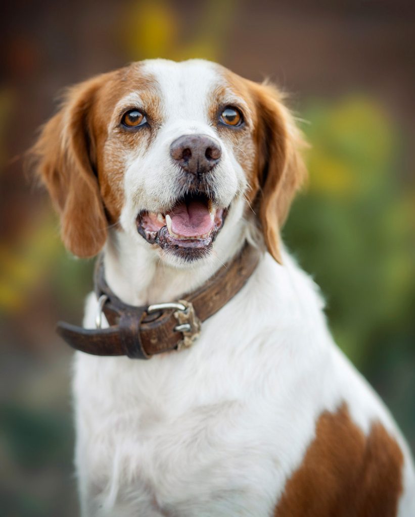
Nala, a senior mongrel dog and beloved family companion, began to show an unusual change three months prior to her veterinary visit. Her nasal planum, normally black in healthy dogs, gradually turned pink. At first, her caregivers assumed the change was temporary. However, when the discolouration persisted and slight ulceration began to appear, they turned to Vetlit Mobile Veterinary Services for answers.
Clinical Examination: Detecting Subtle Clues
On clinical examination, Nala was found to be in good general health, with a body condition score of 4 out of 5. His mucous membranes were pink, his coat was smooth and shiny, and his behavior remained normal. The nasal planum appeared pink and showed signs of ulceration and erosion. There was a colorless, serous nasal discharge present, although the nasal passages were distinct and unobstructed. A whole blood sample was collected and submitted for laboratory analysis, which revealed eosinophilic inflammation, with all the other parameters within normal range, indicating a likely allergic or immune-mediated process affecting the nasal tissues.
Diagnostic Workup: Uncovering the Underlying Cause
A whole blood sample was submitted for laboratory analysis. Results showed eosinophilic inflammation, suggesting an allergic or immune-mediated process. All other blood parameters, including white and red blood cell morphology, were within normal limits.
Based on her clinical presentation and lab findings, our veterinarians made a tentative diagnosis of Discoid Lupus Erythematosus (DLE), a relatively benign but chronic autoimmune skin disease in dogs.
What Is Discoid Lupus Erythematosus (DLE) in Dogs?
DLE is a non-life-threatening autoimmune skin condition in dogs, commonly affecting the nasal planum, and sometimes extending to the eyes, footpads, perianal region, and ears.
Clinical signs include:
- Erythema (redness)
- Depigmentation
- Erosion and ulceration
- Loss of nasal planum texture
- Crusting
The condition is photo-aggravated, meaning sunlight exposure can worsen lesions. That’s why sun protection becomes a key aspect of responsible pet care in dogs with DLE.
Treatment plan: Tailored management for quality of life
Our approach at Vetlit Mobile Veterinary Services focused on reducing sun exposure, symptomatic relief and Immunosuppressive therapy. After one month of treatment, Nala showed remarkable improvement. The ulceration reduced, the depigmentation stabilised, and she resumed her normal daily life.
Takeaway for Dog Owners
Autoimmune conditions like DLE may not be common, but they highlight the importance of early detection, regular check-ups, and responsible dog ownership.